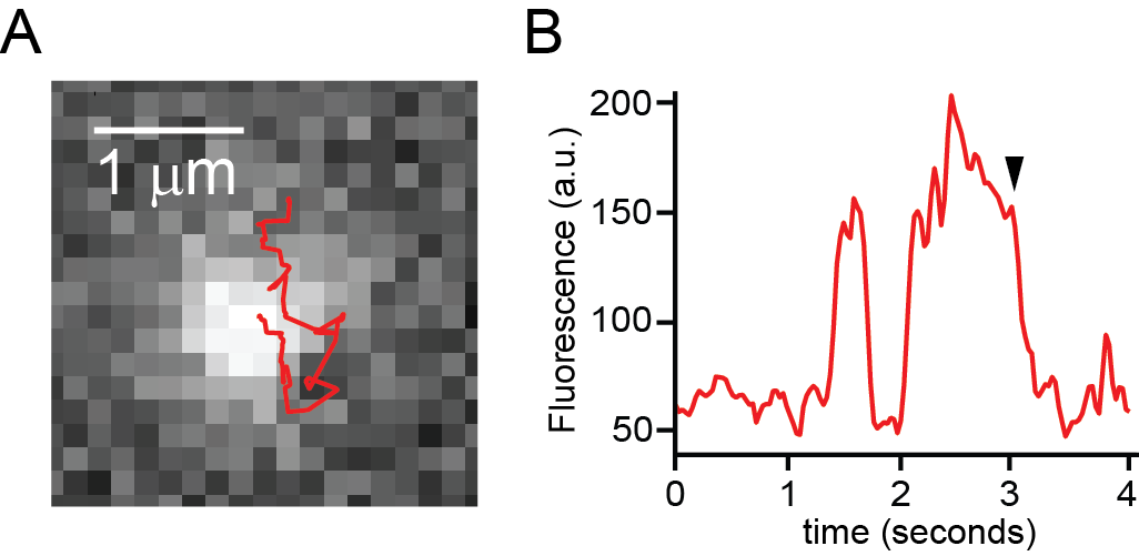
Figure 1. Sparklet
We use fluorescence microscopy and Ca2+ sensitive dyes to record the time-dependent behavior of single TRPV1 ion channels opening and closing in the cell. Our studies revealed that TRPV1 became slower with time that it remained open (Senning et al., eLife 2015). In the figure to the left I observe Ca2+ sparklets overlap with mobile TRPV1-GFP in plasma membrane of a HEK293T /17 cell. (A) Track of TRPV1-GFP with sparklet image as background. Clicking on image provides further examples of sparklets (http://faculty.washington.edu/endsen/Sparklets.m4v). (B) Fluorescence intensity of mobile TRPV1-GFP in (A). Sustained higher intensities correspond to sparklet activity. Arrowhead indicates acquisition of image in (A).
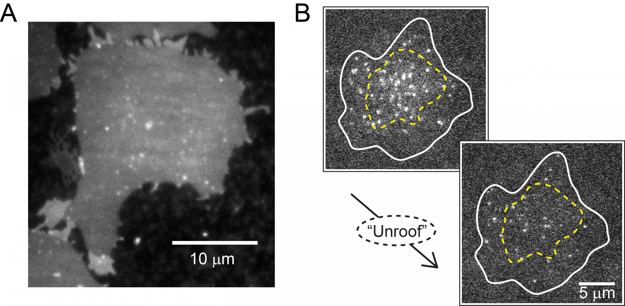
Figure 2. Unroofed cells

Cell unroofing is the process by which we remove the body of an adherent cell grown on a coverslip so that the only remaining cellular material on the coverslip is a plasma membrane sheet. Our recent structural studies of TRPV1 with an unnatural amino acid (UAA) inserted at different sites along its polypeptide sequence relied on sonication as the unroofing method (Zagotta et al., JGP 2016). To the left are examples of unroofed HEK293T/17 cells. (A) A plasma membrane sheet loaded with fluorescent Rhodamine B C-18. Brighter regions on periphery indicate partial unroofing where two bilayers are still present. Clicking here shows an unroofing movie of a cell expressing the PIP2 marker PLCd-PH-GFP. The fluorescence intensity decreases after unroofing as the PLCd-PH-GFP dissociates from the membrane. (B) A cell (outline in white) expressing TRPV1-GFP has a region of diffuse background fluorescence (yellow dashed line). The plasma membrane with resident TRPV1-GFP single channels remains after unroofing while intracellular TRPV1-GFP (presumably in endoplasmic reticulum and other intracellular membranes) is lost (Sponholtz and Senning bioRxiv 2021).
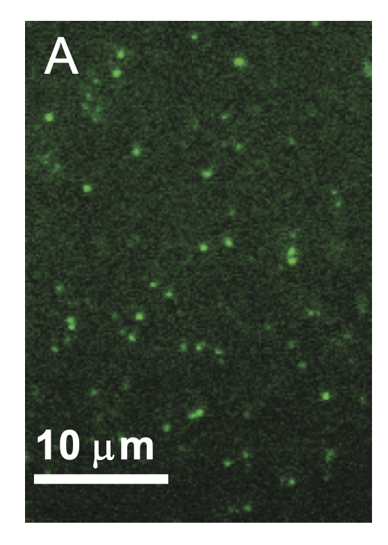
Figure 3.
Single molecule lipid imaging is conducted in unroofed cells with genetically encoded lipid headgroup recognition motifs known as PH domains. The image shows two unroofed cells with PLCdelta1-PH domain labeled GFP bound to various positions on the inner leaflet. The image links to a movie (PLCdelta1-PH-GFP single molecule), showing the highly dynamic character of the labaled PH domains as these bind PI(4,5)P2 lipids and traverse the plasma membrane sheet before dissociating or photobleaching (Sponholtz and Senning bioRxiv 2021).


