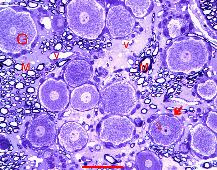
Fig. 5.17. Ganglion nerve cells (G) - a semithin epoxy-resin section. Light microscopy of semithin (0.5 µm) sections from material processed for electron microscopy offers high resolution views intermediate between conventional light microscopy and electron microscopy. These cells belong to the largest cells of the body. M - groups of myelinated axons, V - blood vessels, arrow - satellite cells, empty arrow - nucleus. Scale = 100 µm. (Rat, trigeminal ganglion.)