

Instrumentation


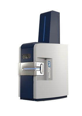
Bruker timsTOF Flex Mass Spectrometer
The Bruker timsTOF Flex Mass Spectrometer allows for both MALDI and Electrospray introduction of samples facilitating both mass spectrometry imaging and traditional LC-MS analysis. It has the added capability of trapped ion mobility which can be used to separate isobaric/isomeric species which may have very different spatial localization. Analytes for fragmentation can be selected based on both their mobility and m/z allowing for much cleaner fragmentation spectra to be obtained, increasing the likelihood of confident identification. It is equipped with a SmartBeam 3D™ laser capable of a laser focus on tissue of <5 µm. This instrment is capable of collecting up to 40 pixels per second during imaging acquistions. Direct software integration with SCiLS Lab and MetaboScape allow for facile identification of metabolites, lipids, and glycans.
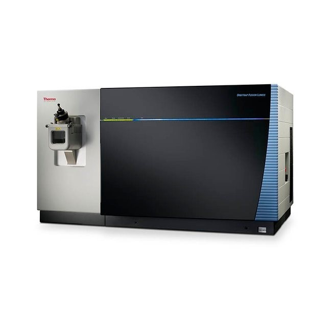
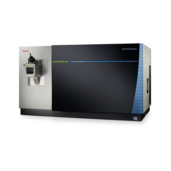
Thermo Fusion Lumos Tribrid Mass Spectrometer
The Thermo Fusion Lumos Tribrid Mass Spectrometer allows for very high resolution and accurate mass measurements of biomolecules. This capability allows for accurate determination of molecular formulas, helping to facilitate analyte identification. The Lumos Fusion is coupled with a MassTech AP/MALDI source for mass spectrometry imaging of lipids and metabolites. Integrated UVPD capabilities aid in biomolecule fragmentation for identification.
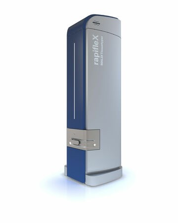
Bruker RapifleX MALDI TOF/TOF Mass Spectrometer
The Bruker RapifleX MALDI TOF/TOF Mass Spectrometer is the premier instrument for high-speed MALDI mass spectrometry imaging. The RapifleX allows for rapid data collection of up to 40 pixels per second. It is equipped with a SmartBeam 3D™ laser capable of a laser focus on tissue of <5 µm. Data are directly co-registered to an optical image of the tissue section allowing for evaluation of biomolecular signals in desired histological features. SCiLS Lab software allows for simultaneous visualization of multiple image files as well as statistical analysis including segmentation, hypothesis testing, and classification algorithm generation. This instrument is on loan from Bruker.
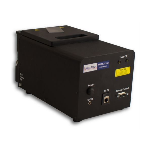
MassTech AP/MALDI (ng) UHR
The MassTech AP/MALDI (ng) UHR is an add-on MALDI source that can be coupled to the Thermo Fusion Lumos mass spectrometer. The tunable Nd:YAG laser with a repetition rate of 10 kHz allows for high throughput mass spectrometry imaging with an achievable spatial resolution of 10 μm. The source operates at atmospheric pressure, as opposed to the high vacuum of traditional MALDI sources, producing somewhat complementary ions and allowing for analysis of samples that are not vacuum compatible.
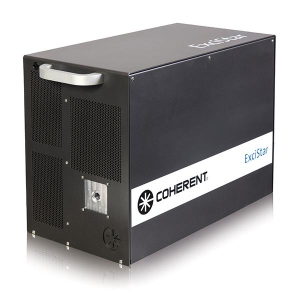
Coherent ExciStar 500 Excimer Laser
The Coherent ExciStar 500 Excimer Laser is an ultraviolet wavelength laser (193 nm) that can be coupled to a mass spectrometer to perform ultraviolet photodissociation (UVPD) of biomolecules in the gas phase to aid in characterization and identification. The laser operates at a repetition rate up to 500 Hz with a tunable pulse energy up to 5 mJ. UVPD produces complementary fragmentation to other MS/MS techniques, often keeping posttranslational modifications of proteins intact and allowing for localization of double bonds in lipid fatty acids. The ExciStar 500 Laser is currently coupled to the Thermo Fusion Lumos mass spectrometer.
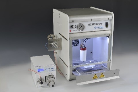
HTX M5 Robotic Reagent Sprayer
The HTX M5 Robotic Reagent Sprayer allows for high-throughput, reproducible sample preparation for MALDI analysis. Reagents such as derivatization agents, proteolytic enzymes, and/or MALDI matrices can be deposited in a uniform manner over tissue sections. Fine control of flow rate, temperature, gas pressure, and nozzle track speed allow for the generation of very small droplets (<5 µm), minimizing delocalization of analytes and allowing for high spatial resolution imaging.
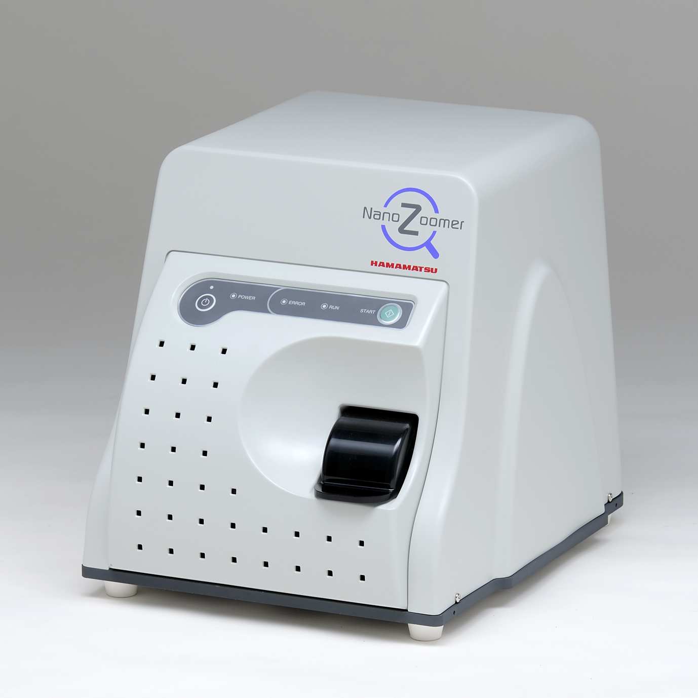
Hamamatsu NanoZoomer SQ
The Hamamatsu NanoZoomer SQ is a high-quality compact digital whole slide scanner. It is capable of scanning a 15x15 mm area at 20x magnification in about 2.5 minutes. It produces seamless brightfield microscopy images that can be directly imported into MSI analysis software. Images can be reviewed and annotated using the NDP.view2 viewer software for region of interest analysis from MSI data.
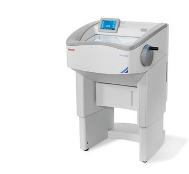
Thermo NX50 Cryostat
The Thermo NX50 Cryostat allows for high-quality, reproducible, sectioning of frozen tissue specimens. Sectioning is the first step in sample preparation for mass spectrometry imaging and obtaining quality sections is of utmost importance in generating optimal image data. The cryostat allows for section thickness from 0.5 to 100 µm and an operating temperature range from -10 to -43°C. Specimen head alignment minimizes sample loss with repeated sectioning of a block. Adjustable height of the system allows for ergonomic sectioning for all users.