

Tutorial Presentations
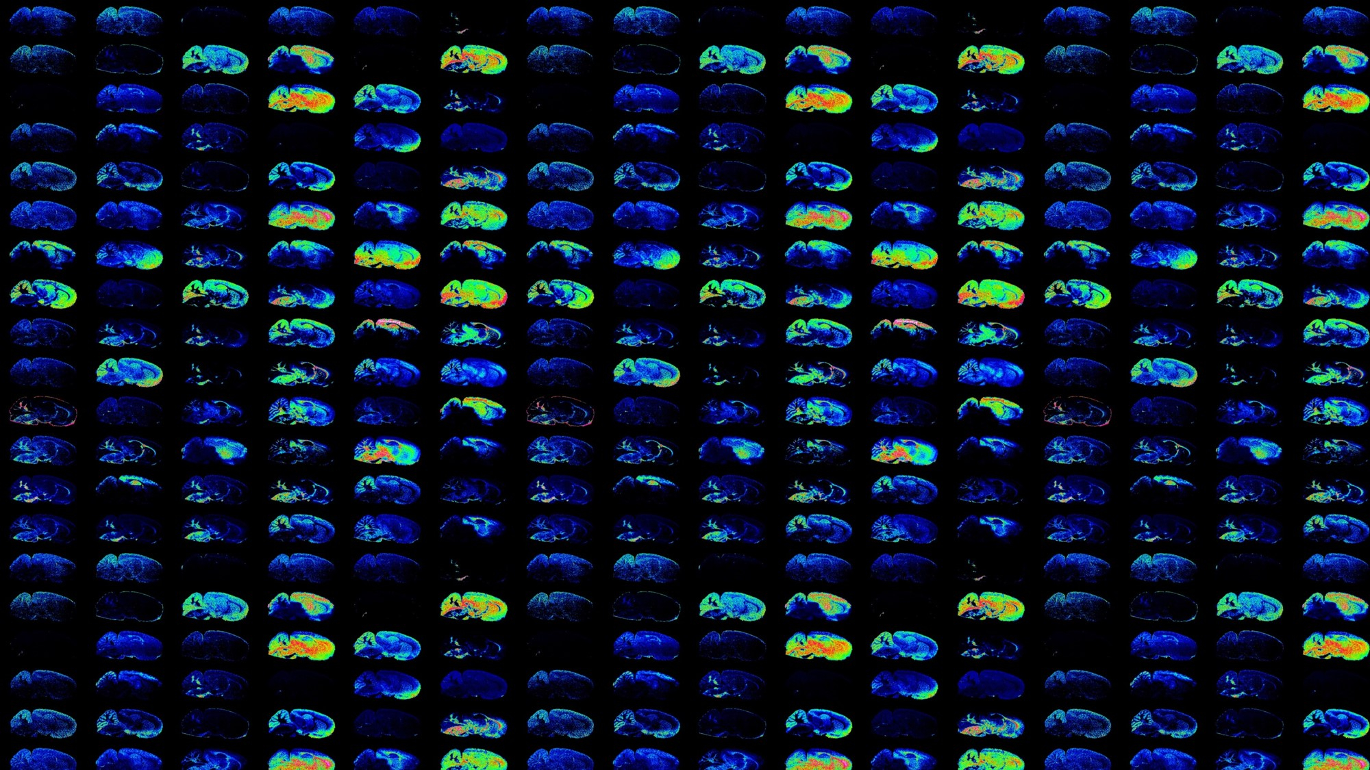 Mass Spectrometry Imaging Overview
Mass Spectrometry Imaging (MSI) provides a molecular snapshot of biological processes going on in a tissue specimen. Thin tissue sections are analyzed in an ordered array over the surface of the sample using a laser or solvent spray to desorb and ionize molecules that can be subsequently detected in a mass spectrometer. Hundreds to thousands of biomolecules can be detected from a single tissue section without prior knowledge of the molecules present or the need for target specific reagents, making MSI an ideal technology for biomarker discovery.
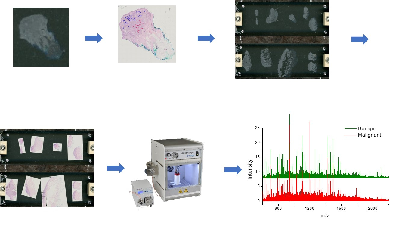
Histology-Guided Mass Spectrometry Profiling Overview
The Histology-Guided Mass Spectrometry (HGMS) Profiling workflow is used for high-throughput analysis of clinical samples. This approach allows for highly targeted analysis of specific cell types or histological features through the use of histological staining to guide data collection. HGMS data is highly conducive to statistical analysis and classification algorithm generation.
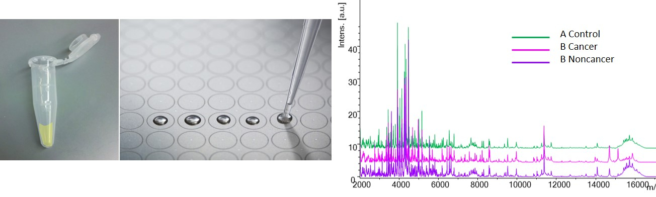
Biofluid/Cytology Profiling Overview
Biofluids, scrapes, lavages, and other minimally invasive collection techniques are often used for diagnostic or prognostic testing. These samples are also able to be analyzed by mass spectral profiling. It is important, however, that the sample is closely related to the disease being studied as dilution effects can hinder the success of the analysis.
Clinical MSI Practical Considerations
This powerpoint presentation video outlines practical consideration for clinical MSI studies. Included are discussions of experimental planning, laboratory and instrumentation considerations, needs for statistical analysis, and an example of a clinical MSI study. This video should be helpful for clinicians thinking about doing a collaborative MSI study as well as researchers planning to do a clinical MSI study in their own lab. A pdf of the slides is also available for download.