Rossi RM, Yum L, Agaisse H, Payne SM.
Cardiolipin Synthesis and Outer Membrane Localization Are Required for Shigella flexneri Virulence. mBio [Internet]. 8 (4).
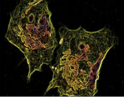
Publisher's VersionAbstractCardiolipin, an anionic phospholipid that resides at the poles of the inner and outer membranes, is synthesized primarily by the putative cardiolipin synthase ClsA in Shigella flexneri. An S. flexneri clsA mutant had no cardiolipin detected within its membrane, grew normally in vitro, and invaded cultured epithelial cells, but it failed to form plaques in epithelial cell monolayers, indicating that cardiolipin is required for virulence. The clsA mutant was initially motile within the host cell cytoplasm but formed filaments and lost motility during replication and failed to spread efficiently to neighboring cells. Mutation of pbgA, which encodes the transporter for cardiolipin from the inner membrane to the outer membrane, also resulted in loss of plaque formation. The S. flexneri pbgA mutant had normal levels of cardiolipin in the inner membrane, but no cardiolipin was detected in the outer membrane. The pbgA mutant invaded and replicated normally within cultured epithelial cells but failed to localize the actin polymerization protein IcsA properly on the bacterial surface and was unable to spread to neighboring cells. The clsA mutant, but not the pbgA mutant, had increased phosphatidylglycerol in the outer membrane. This appeared to compensate partially for the loss of cardiolipin in the outer membrane, allowing some IcsA localization in the outer membrane of the clsA mutant. We propose a dual function for cardiolipin in S. flexneri pathogenesis. In the inner membrane, cardiolipin is essential for proper cell division during intracellular growth. In the outer membrane, cardiolipin facilitates proper presentation of IcsA on the bacterial surface.
Peng ED, Payne SM.
Vibrio cholerae VciB Mediates Iron Reduction. Journal of Bacteriology [Internet]. 199 (12).
Publisher's VersionAbstractVibrio cholerae is the causative agent of the severe diarrheal disease cholera. V. cholerae thrives within the human host, where it replicates to high numbers, but it also persists within the aquatic environments of ocean and brackish water. To survive within these nutritionally diverse environments, V. cholerae must encode the necessary tools to acquire the essential nutrient iron in all forms it may encounter. A prior study of systems involved in iron transport in V. choleraerevealed the existence of vciB, which, while unable to directly transport iron, stimulates the transport of iron through ferrous (Fe2+) iron transport systems. We demonstrate here a role for VciB in V. cholerae in which VciB stimulates the reduction of Fe3+ to Fe2+, which can be subsequently transported into the cell with the ferrous iron transporter Feo. Iron reduction is independent of functional iron transport but is associated with the electron transport chain. Comparative analysis of VciB orthologs suggests a similar role for other proteins in the VciB family. Our data indicate that VciB is a dimer located in the inner membrane with three transmembrane segments and a large periplasmic loop. Directed mutagenesis of the protein reveals two highly conserved histidine residues required for function. Taken together, our results support a model whereby VciB reduces ferric iron using energy from the electron transport chain.