Milich KM, Koestler BJ, Simmons JH, Nehete PN, Di Fiore A, Williams LE, Dudley JP, Vanchiere J, Payne SM.
Methods for detecting Zika virus in feces: A case study in captive squirrel monkeys (Saimiri boliviensis boliviensis). PLoS One [Internet]. 13 (12).
Publisher's VersionAbstractA strain of Zika virus (ZIKV) of Asian origin associated with birth defects and neurological disorders has emerged and spread through the Americas. ZIKV was first isolated in the blood of nonhuman primates in Africa and has been detected in the blood, saliva, and urine of a few catarrhine species in both Africa and Asia, suggesting that nonhuman primates may serve as both a source and a reservoir of the virus. The recent introduction of ZIKV to human populations in the Americas presents the potential for the virus to spread into nonhuman primate reservoirs. Thus, it is critical to develop efficient and noninvasive detection methods to monitor the spread of the virus in wild nonhuman primate populations. Here, we describe a method for ZIKV detection in noninvasively collected fecal samples of a Neotropical primate. Fecal samples were collected from two captive squirrel monkeys (Saimiri boliviensis boliviensis) that were experimentally infected with ZIKV (Strain Mexico_1_44) and an additional two uninfected squirrel monkeys. Nucleic acids were extracted from these samples, and RT-qPCR was used to assay for the presence of ZIKV using primers flanking a 101 bp region of the NS5 gene. In both ZIKV-inoculated animals, ZIKV was detected 5-11 days post-infection, but was not detected in the uninfected animals. We compare the fecal results to ZIKV detection in serum, saliva, and urine samples from the same individuals. Our results indicate that fecal detection is a cost-effective, noninvasive method for monitoring wild populations of Neotropical primates as possible ZIKV reservoirs.
Koestler BJ, Fisher CR, Payne SM.
Formate Promotes Shigella Intercellular Spread and Virulence Gene Expression. mBio [Internet]. 9 (5) :e01777-18.
Publisher's VersionAbstract
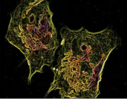
The intracellular human pathogen Shigella flexneri invades the colon epithelium, replicates to high cell density within the host cell, and then spreads to adjacent epithelial cells. When S. flexneri gains access to the host cytosol, the bacteria metabolize host cytosolic carbon using glycolysis and mixed acid fermentation, producing formate as a by-product. We show that S. flexneri infection results in the accumulation of formate within the host cell. Loss of pyruvate formate lyase (PFL; ΔpflB), which converts pyruvate to acetyl coenzyme A (CoA) and formate, eliminates S. flexneri formate production and reduces the ability of S. flexneri to form plaques in epithelial cell monolayers. This defect in PFL does not decrease the intracellular growth rate of S. flexneri; rather, it affects cell-to-cell spread. The S. flexneri ΔpflB mutant plaque defect is complemented by supplying exogenous formate; conversely, deletion of the S. flexneri formate dehydrogenase gene fdnG increases host cell formate accumulation and S. flexneri plaque size. Furthermore, exogenous formate increases plaque size of the wild-type (WT) S. flexneristrain and promotes S. flexneri cell-to-cell spread. We also demonstrate that formate increases the expression of S. flexneri virulence genes icsA and ipaJ Intracellular S. flexneriicsA and ipaJ expression is dependent on the presence of formate, and ipaJ expression correlates with S. flexneri intracellular density during infection. Finally, consistent with elevated ipaJ, we show that formate alters S. flexneri-infected host interferon- and tumor necrosis factor (TNF)-stimulated gene expression. We propose that Shigella-derived formate is an intracellular signal that modulates virulence in response to bacterial metabolism.
IMPORTANCEShigella is an intracellular pathogen that invades the human host cell cytosol and exploits intracellular nutrients for growth, enabling the bacterium to create its own metabolic niche. For Shigella to effectively invade and replicate within the host cytoplasm, it must sense and adapt to changing environmental conditions; however, the mechanisms and signals sensed by S. flexneri are largely unknown. We have found that the secreted Shigella metabolism by-product formate regulates Shigella intracellular virulence gene expression and its ability to spread among epithelial cells. We propose that Shigella senses formate accumulation in the host cytosol as a way to determine intracellular Shigella density and regulate secreted virulence factors accordingly, enabling spatiotemporal regulation of effectors important for dampening the host immune response.
Koestler BJ, Ward CM, Payne SM.
Shigella Pathogenesis Modeling with Tissue Culture Assays. Current Protocols in Microbiology [Internet]. 50 (1) :e57.
Publisher's VersionAbstractShigella is an enteroinvasive human pathogen that infects the colonic epithelium and causes Shigellosis, an infectious diarrheal disease. There is no vaccine for the prevention or treatment of Shigellosis and antibiotic‐resistant strains of Shigella are increasing, emphasizing the need for a deeper understanding of Shigella pathogenesis in order to design effective antimicrobial therapies. Small animal models do not recapitulate Shigellosis, therefore tissue‐cultured cells have served as model systems to study Shigellapathogenesis. Here, protocols to enumerate Shigella invasion, cell‐cell spread, and plaque formation in the tissue‐cultured cell lines Henle‐407 and CoN‐841 are described. Additionally, a new method to study Shigella invasion in primary intestinal enteroids is described. These protocols can be used to examine different aspects of Shigella virulence.